Researchers Work to See the Heart More Clearly and Save Lives
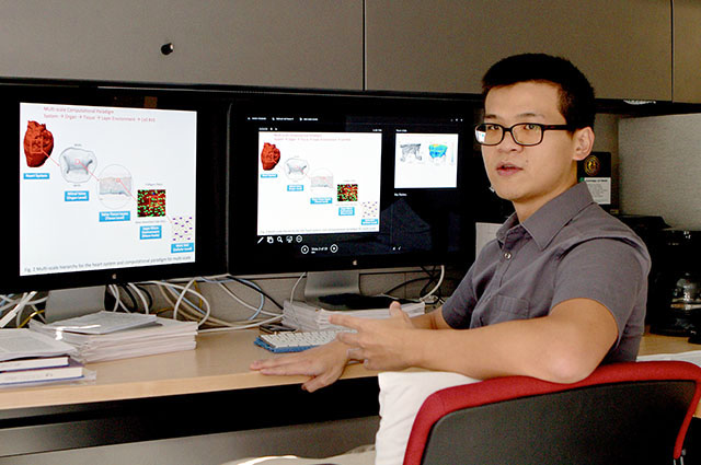
Postdoctoral fellow Chung-Hao Lee and colleagues at the University of Texas are mapping the heart’s mitral valve to make surgery safer. Photo by Andrew Masi.
By Reihaneh Hajibeigi
For Reporting Texas
“That’s your mitral valve,” said postdoctoral fellow Chung-Hao Lee, pointing to a 3D simulation that fills his computer screen with a pair of lips that flap open and then close, a rainbow of colors indicating how tightly. The display lies at the core of work by Lee and his colleagues at the department of biomedical engineering at the University of Texas at Austin to improve the visibility of one of the heart’s trickiest pieces of real estate.
The difficulty of seeing the mitral valve clearly, in its natural, functioning state, creates complications for heart surgeons who, according to the Texas Heart Institute, repair the organ some 99,000 times a year in the United States at an average cost of $160,000 per procedure.
Eliminating such blind spots is vital. The mitral valve, Lee said, “is what controls the one-directional blood flow from the left atrium to the left ventricle, and if it doesn’t close properly,” the malfunction can lead to heart failure, stroke, blood clots and, if not treated properly, death.
Under the direction of biomedical engineering professor Michael Sacks, the UT-Austin research team hopes its simulation work will let practitioners to stop mentally imaging the mitral valve and instead view real-time images on a computer screen.
Having that option is critical during surgery, when doctors reroute the patient’s blood flow and the mitral valve collapses like a spent parachute. Surgeons now rely on experience to recreate the valve’s shape in their mind’s eye. That lack of precision can lead to costly follow-up surgeries, some 40,000 in the United States each year.
Sacks and his team use micro-CT images of the valve at different blood pressures to build the right valve geometry for a generic simulation. Micro-CT technology allows researchers to layer large numbers of X-ray images into 3D models without affecting the original, in this case the human heart. Ultimately, the UT-Austin team aims to tailor simulations for individual patients.
In September, the National Institutes of Health awarded the project a $6.6 million grant, which includes UT-Austin and also employs in-vitro measurements of the mitral valve from the Georgia Institute of Technology and animal studies from the University of Pennsylvania.
Sacks and his team are developing and testing current solutions for mitral valve repair, including commonly used annuloplasty rings. Surgical repair typically involves implanting this circular device around the mitral valve, which then pulls the leaflets of the valve together to re-establish the proper shape and function.
Annuloplasty rings are usually made of plastic, silicone or metal, and researchers hope to use the simulation technology to test which material puts the least stress on mitral valve tissues after implantation.
“Our research will tell us how to augment the shape of the valve post-surgery and how the cells in the tissue will respond to the stresses,” said Lee.
Sacks, who directs the Center for Cardiovascular Simulation in the Institute of Computational Engineering and Sciences, said he believes the details of the model, from the cells all the way up to the entire organ, will allow the mechanics of the whole system to be analyzed.
“We hypothesize the reason the mitral valve fails is because of how the abnormal stresses affect the underlying cells that lie within the mitral valve tissues,” Sacks said. “It’s not the cells that fail; it’s the organ, so creating the whole mitral valve will let us analyze the whole system.”
Once Sacks and his team have constructed a reliable computational model, they will test the simulations in a clinical setting. The simulations will be patient-specific and allow cardiologists to see the living valve, even when there is no blood flowing through it.
The goal for Sacks and collaborators is designs that are ready for clinical trials by 2018, when the current five-year grant ends.
“This will hopefully be used as a clinical tool to provide certain information to surgeons in order to decide which strategy is best for each patient,” Lee said. “Eventually, we will be able to apply a particular patient’s valve conditions to the simulation and figure out the best solution for them.”
John Barnard, assistant clinical professor at Weill Cornell Medical College in New York, said the mitral valve is a 3D structure, so creating a 3D image will give cardiologists a better idea of which part needs to be fixed.
“The cardiology field relies on images more and more,” said Barnard, a 30-year veteran as a cardiologist. “When a surgeon is operating, the valve is not at the shape it is when the body is working, so creating these simulations showing how the valve looks on a day-to-day basis will allow doctors to be even more prepared prior to surgery.”